
.webp)
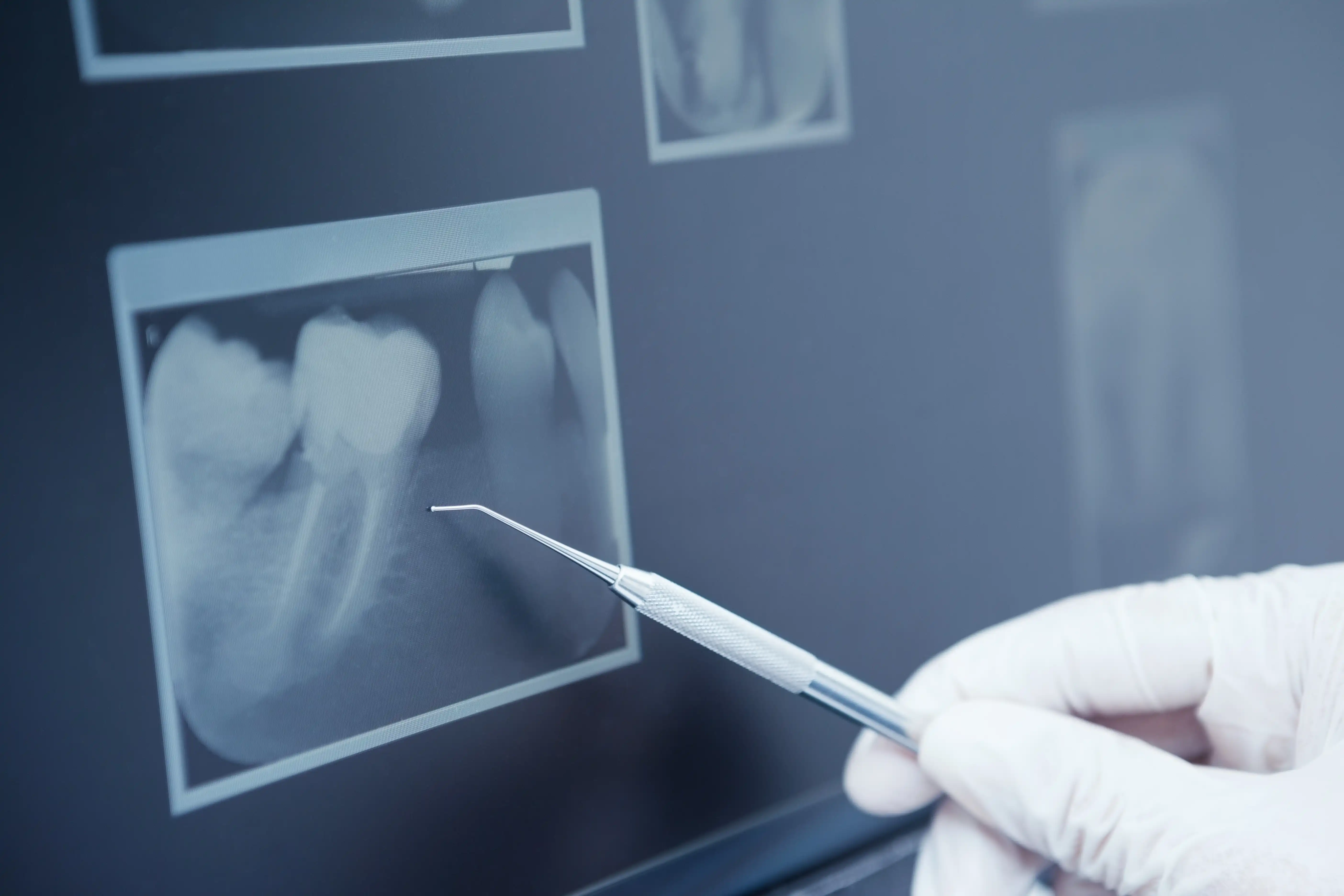
The most common type taken at routine exams. Shows the upper and lower back teeth simultaneously, revealing cavities forming between teeth and early signs of bone loss from gum disease that are impossible to see during a visual exam.
Dental X-Rays and Imaging in Richmond, TX
Digital Imaging by Dr. William Sung DMD
.webp)
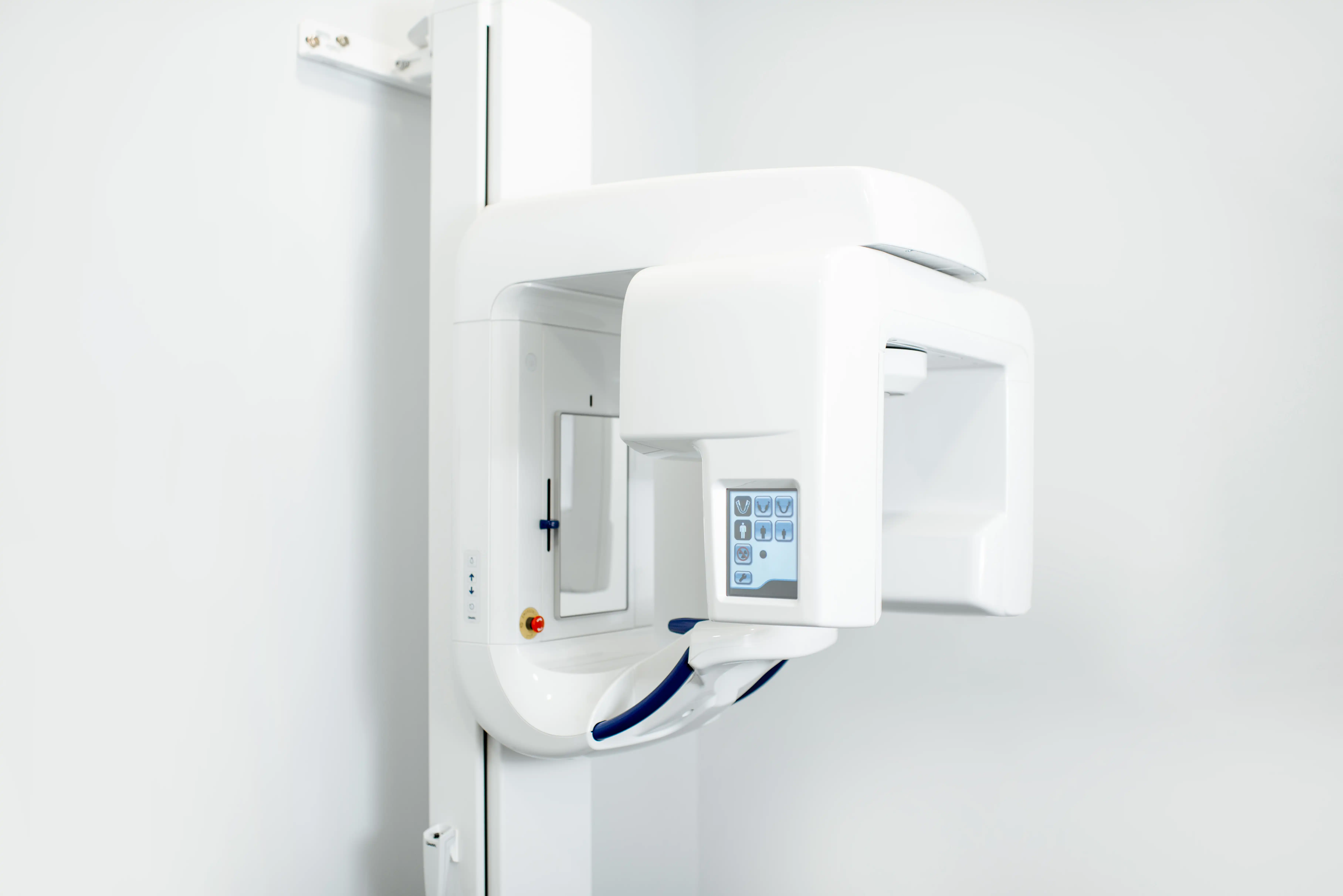
Dental X-Rays and Imaging at Grand Mission
Some of the most important things happening in your mouth are completely invisible to the naked eye. Cavities forming between teeth, bone loss beneath the gumline, infections developing at the root, cysts, and early-stage tumors all appear on X-rays long before they cause symptoms you would notice. At Grand Mission, Dr. William Sung uses digital X-ray technology and 3D cone beam CT imaging to see the full picture of your oral health quickly, safely, and with far less radiation than traditional film-based systems.
What Dental X-Rays Are Used For
Different types of imaging reveal different things. Here is what each type is designed to detect.
Shows the entire tooth from crown to root tip and the surrounding bone. Used to evaluate a specific tooth when there is pain, sensitivity, or a suspected infection at the root.
A single wide-view image showing all teeth, the jaw, sinuses, and surrounding structures. Used for treatment planning, evaluating wisdom teeth, and assessing the full scope of bone levels and dental development.
Cone Beam CT scanning produces a precise three-dimensional map of your teeth, bone, nerves, and sinuses. Used for implant planning, bone grafting, and any case where precise anatomical detail determines the outcome of treatment.
What to Expect During Dental Imaging
Sensor or Film Placement
A small digital sensor is positioned inside your mouth to capture the target area. The process is quick and comfortable for the vast majority of patients.
Image Capture
The X-ray is taken in a fraction of a second. Digital sensors capture the image instantly and display it on screen within seconds, allowing Dr. Sung to review and discuss findings with you immediately.
AI-Assisted Analysis
Grand Mission uses an FDA-validated AI diagnostic platform to assist in analyzing X-ray images, helping identify areas of concern with a level of precision that goes beyond visual review alone. This technology adds a layer of diagnostic accuracy to every image taken.
Review and Explanation
Dr. Sung walks you through exactly what your images show in plain language. No jargon, no assumptions. You understand your oral health fully before any treatment decision is made.
What Sets Grand Mission Apart
Grand Mission uses digital X-rays that produce up to 90% less radiation than traditional film-based systems, paired with 3D cone beam CT imaging for complex cases and an FDA-validated AI diagnostic platform that adds precision to every image analyzed.
Digital X-Rays with Up to 90% Less Radiation
Our digital imaging system captures clearer, more detailed images than traditional film while exposing patients to significantly less radiation. Images appear instantly on screen so Dr. Sung can review findings with you during your appointment.
.webp)
CBCT 3D Imaging for Complex Cases
When implant placement, bone grafting, or complex treatment planning is involved, Grand Mission uses cone beam CT scanning to produce a precise three-dimensional map of your anatomy before any procedure begins.
AI-Assisted Screening Technology
An FDA-validated AI platform assists in analyzing every X-ray taken at Grand Mission, identifying areas of concern with a level of precision that supports Dr. Sung's clinical judgment at every exam.
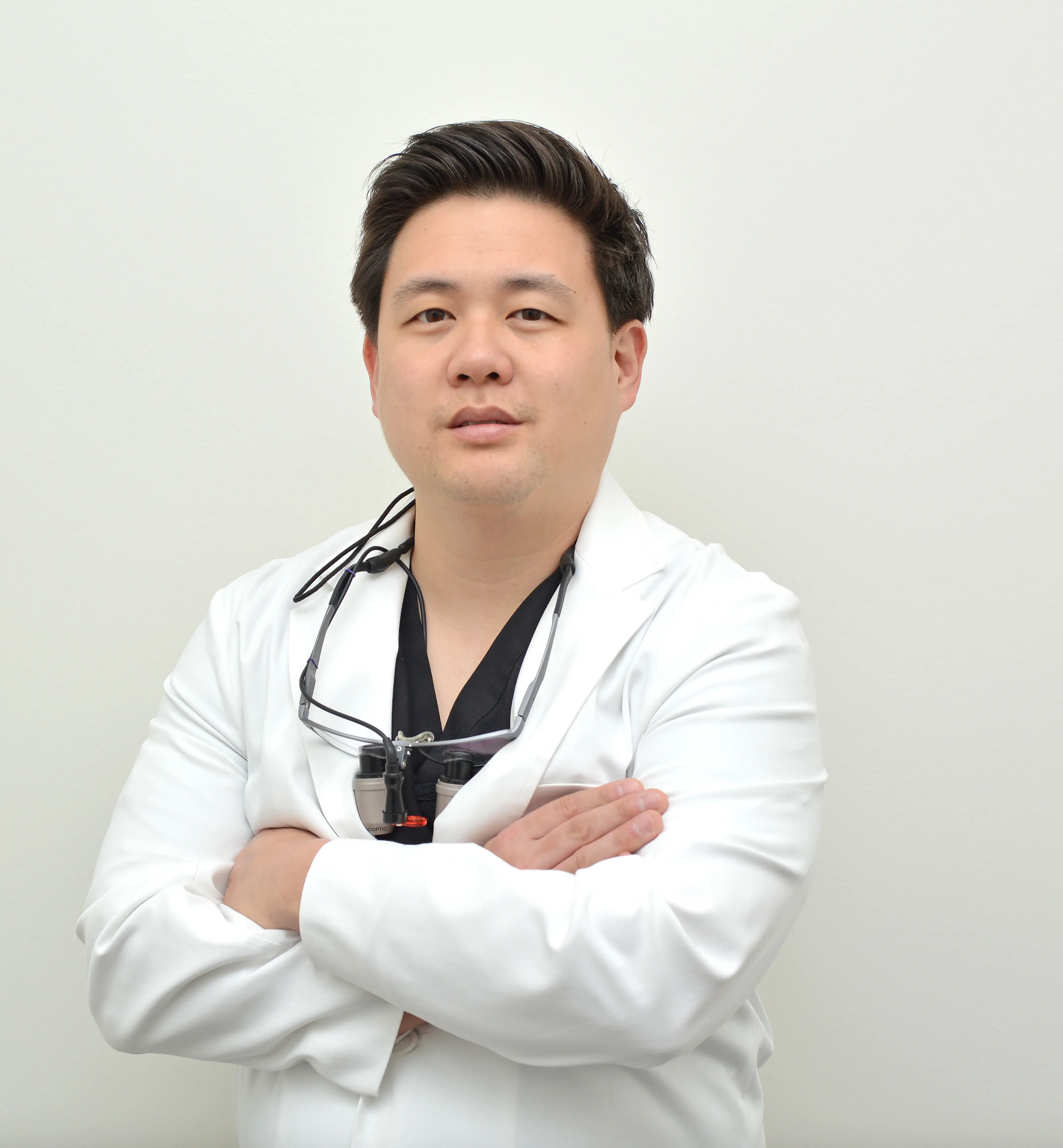
Dr. William Sung DMD

Dr. Tam Nguyen DMD
.webp)
Dr. Van Nguyen DDS

New Patient Special!
Starting at
$189
get
Comprehensive exam
X-Rays
Cleaning
Full Treatment Plan
(Cannot be combined with insurance, new patients only, not valid in cases of periodontal disease.)
.webp)
FREE Second Opinion
Special Price
FREE
get
No cost, no pressure — get an honest evaluation without feeling rushed or sold to
Clear answers you can trust — we review your diagnosis, options, and next steps
Confidence before treatment — make informed decisions about your dental health
Frequently Asked Questions About dental X-rays
Are dental X-rays safe?
Yes. Digital X-rays at Grand Mission use up to 90% less radiation than traditional film systems. The dose from a full set of dental X-rays is lower than the radiation exposure from a short flight. We also use lead aprons for every patient as a standard precaution.
How often do I need dental X-rays?
For most patients, bitewing X-rays are taken once a year and a full set of X-rays every three to five years. Patients with a history of decay, gum disease, or active treatment may need imaging more frequently. Dr. Sung recommends only what your specific situation requires.
What is the difference between regular X-rays and a CBCT scan?
Standard dental X-rays produce flat two-dimensional images. CBCT scanning produces a full three-dimensional model of your teeth, bone, nerves, and sinuses. CBCT is used for implant planning, bone grafting, and any case where precise three-dimensional anatomy determines the outcome of treatment.
Do I need X-rays at every appointment?
Not necessarily. Routine bitewing X-rays are taken once a year for most patients. Full mouth X-rays are taken less frequently. CBCT is only used when a specific treatment plan requires it. Dr. Sung will always explain why a specific type of imaging is recommended before proceeding.
What does an AI diagnostic platform do with my X-rays?
The AI platform analyzes your digital X-ray images to flag areas that may indicate early decay, bone changes, or other concerns. It works alongside Dr. Sung's clinical judgment, not instead of it, adding a layer of precision that improves diagnostic accuracy for every patient.


What Dental Imaging Supports
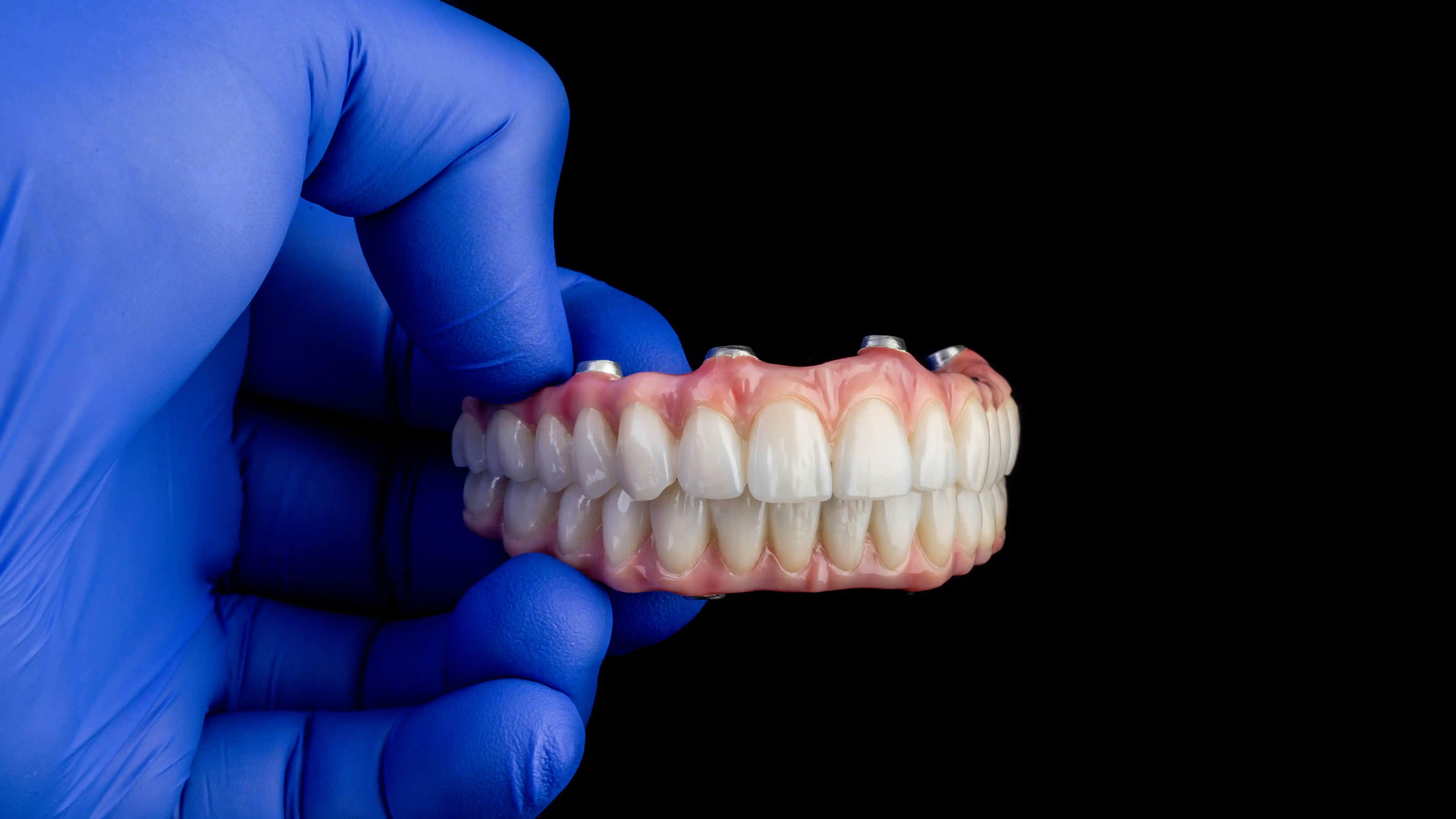
Dental Implants
Description: CBCT 3D imaging is used for every implant case at Grand Mission to map bone density, nerve locations, and jaw anatomy before placement begins.
Learn More about Dental Implants

Bone Grafting
Precise 3D imaging determines exactly how much bone needs to be rebuilt, in which dimensions, and which grafting technique will achieve the best result.
Learn More about Bone Grafting

Oral Cancer Screening
Digital X-rays and AI-assisted analysis complement the visual oral cancer screening performed at every exam, ensuring nothing beneath the surface is missed.
Learn More about Oral Cancer Screening
Your confident smile
Is our guide
Schedule your appointment today
Seeing the full picture is how we catch problems early, plan treatment accurately, and keep your oral health on track for life. Book your exam today and experience the difference that precision imaging makes.